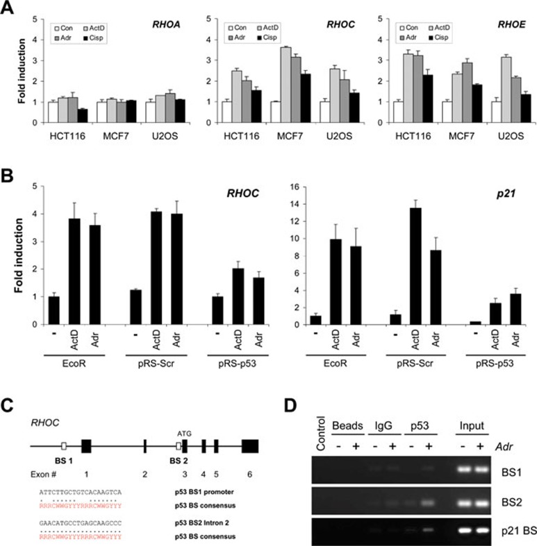
Figure 2.
RHOC is p53 target gene. (A) Genotoxic stress induces RHOC expression. HCT116, MCF-7 or U2OS cells were treated with ActD (2 nM), Adr (0.2 μg/ml) or cisplatin (Cisp; 25 μM) for 24 h. RHOA, RHOC and RHOE mRNA levels were determined by qPCR. Data are presented as mean fold induction ± SEM (n = 3) relative to vehicle control (Con) in each cell line. (B) RHOC induction by genotoxic stress is p53 dependent. Parental MCF-7-Eco, control MCF-7-pRS-Scr and p53-knockdown MCF-7-pRS-p53 cells were treated with ActD (2 nM) or Adr (0.2 μg/ml) for 24 h. RHOC and p21 mRNA levels were determined by qPCR. Data are presented as mean fold induction ± SEM (n = 3), relative to vehicle control. (C) Schematic diagram of the human RHOC gene showing potential p53-binding sites (BS). The p53MH algorithm identified p53-binding sites within the upstream regulatory region (BS1) and the second intron (BS2) of the human RHOC gene. Within each binding site, the individual half-sites are compared with the consensus p53-binding site sequence, where R = purine, Y = pyrimidine and W = adenine or thymine. (D) p53 binds the BS2 element within RHOC intron 2. Chromatin immunoprecipitation was carried out with anti-p53 antibody (DO-7) on chromatin isolated from MCF-7 cells treated with and without Adr (0.2 μg/ml) for 8 h. Ethidium bromide-stained agarose gels show PCR products of RHOC BS2 after immunoprecipitation. Control immunoprecipitations were carried out with beads alone (Beads) or with control mouse IgG (IgG). Input represents 0.5% of the total chromatin used in each condition.

