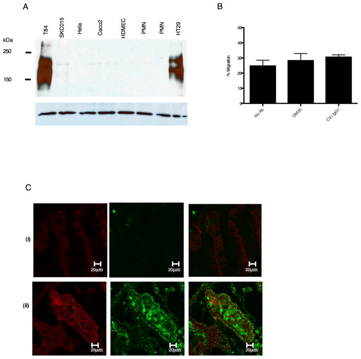
Figure 3. GM35 recognizes an IFNγ responsive glycoprotein.
(A) Following cell lysis, equal amounts of proteins from indicated cells were subjected to SDS-PAGE under reducing conditions and Western blotted with GM35. Data depicts representative results from n=3 Western Blots. (B) Confluent Caco2 monolayers were pre-treated apically with 10 μg/ml GM35 mAb (GM35) before 1×10⁶ PMNs were added to the basolateral surface. PMNs were allowed to migrate in the physiologically relevant basolateral to apical direction for 1 hour in response to a 100nM gradient of n-formyl-methionyl-leucyl-phenylalanine (fMLF). The number of migrated PMNs was quantified by myeloperoxidase assay. Data are means +/− SE (n=3). (C) Cryosections of non-inflamed sections of colonic mucosa (i) and inflamed sections of colonic mucosa (ii) from patients with active ulcerative colitis were examined for localization of the GM35 antigen (green) or the epithelial protein desmoglein (red) as described in methods.

