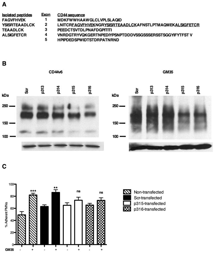
Figure 4. Identification of the GM35 antigen as CD44.
(A) 500cm2 of T84 cells were lysed, immunoprecipitated with GM35 and subjected to tryptic digestion and microsequence analysis. Isolated tryptic peptides mapped to CD44 are underlined. (B) Representative immunoblots demonstrating that transfection of HT29 cells with CD44 gene silencing plasmids (p315, p316) decreased the expression of CD44v6 and the GM35 antigen as compared to scramble control (Scr). (C) HT29 cells were transfected with the same plasmids before measurement of neutrophil adhesion levels. Data are presented as mean ± SE (n=4). Significance was defined at p<0.05 (*, p<0.05; **, p<0.01; ***, p<0.001).

