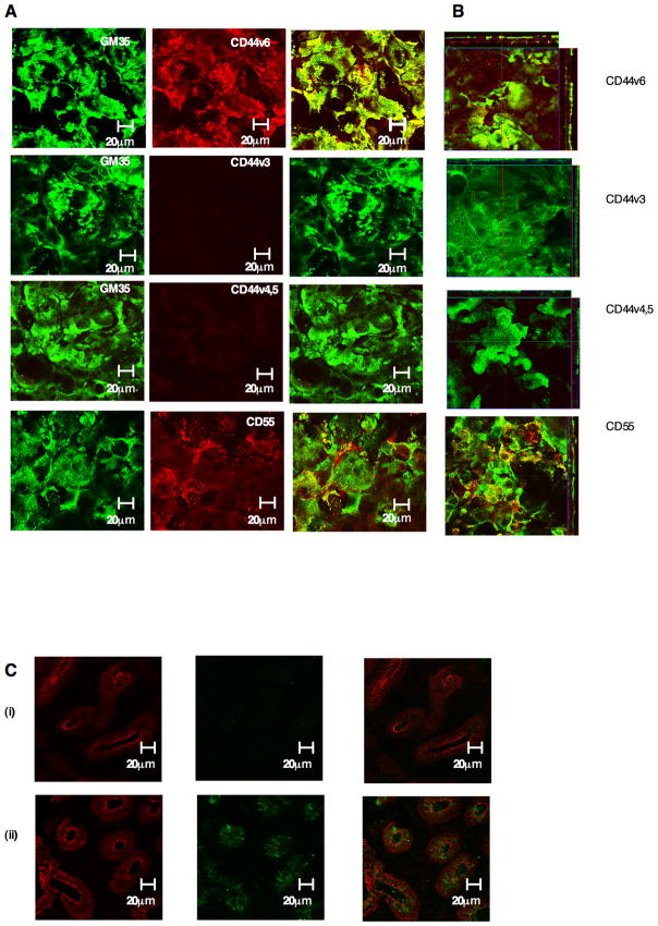
Figure 6. GM35 co-localizes apically with CD44v6.
(A) Non-permeabilized T84 monolayers were co-stained with 10μg/ml Zenon-labeled GM35 (green) and 5μg/ml anti-CD44 antibodies specific for CD44v3, CD44v4,5, CD44v6 or 5μg/ml anti-CD55 mAb. (red). Apical protein localization was determined by confocal microscopy. Images shown are en face (A) or in the x–z plane of section (B). (C) Cryosections of non-inflamed sections of colonic mucosa (i) and inflamed sections of colonic mucosa (ii) from patients with active ulcerative colitis were examined for localization of CD44v6 (Green) or the epithelial marker Desmoglein (red) as described in methods.

