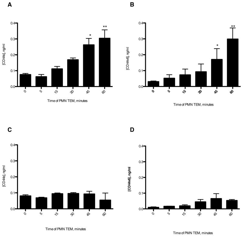
Figure 7. GM35 blocks release of soluble CD44v6.
1×10⁶ PMNs were added to confluent T84 monolayers treated apically with 10μg/ml binding control IgG1 (A,B) or 10 μg/ml GM35(C,D). PMNs were allowed to migrate in the basolateral to apical direction in response to a 100nM gradient of fMLF. At the time- points indicated the T84 coated filters were moved into a fresh fMLF-containing well of a 24-well tissue culture plate. For each indicated time-point the solution from the apical migration reservoir was tested for the presence of soluble CD44s (A,C) or soluble CD44v6 (B,D) by ELISA as described in Methods. Data depicts the mean ± SEM from 3 independent experiments. Significance was defined at p<0.05 (*, p<0.05; **, p<0.01; ***, p<0.001).

