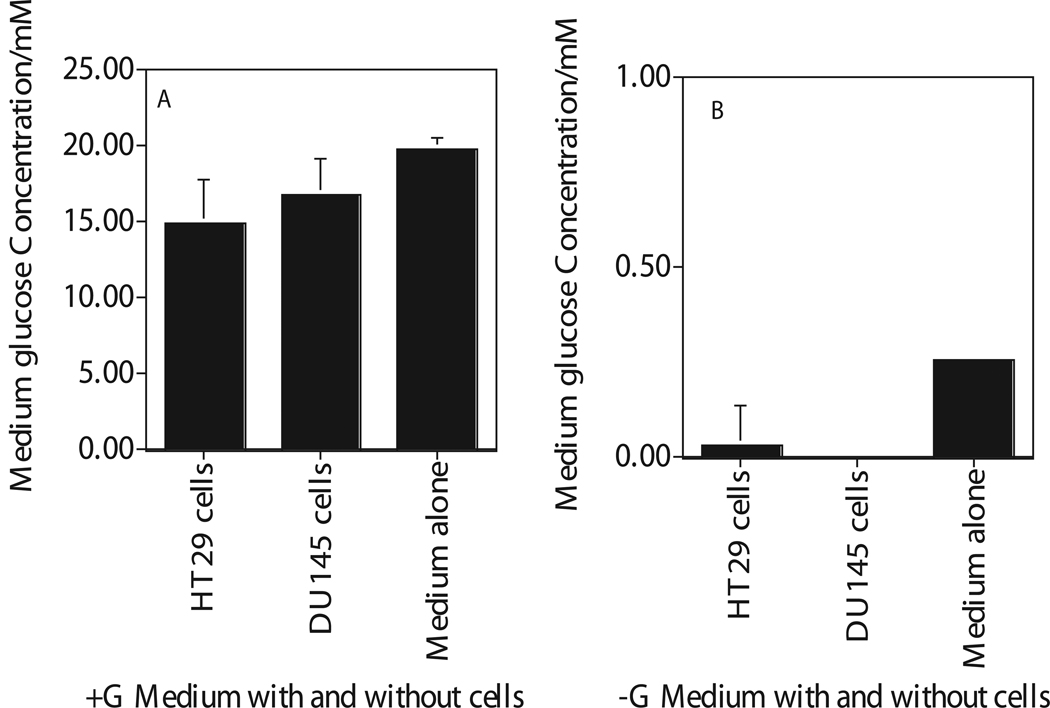
Figure 1.
Enzymatic quantification of extracellular glucose in the medium removed after 4 hours incubation of human cancer cells (HT29, DU145) in glucose (Figure A) and glucose free (Figure B) medium. Medium with Glucose, +G; Medium with zero glucose, −G. Each data point plotted was the mean ± standard error (SE) of at least three experiments with SE as shown unless smaller than points plotted. Statistical comparisons for glucose levels in glucose free medium relative to glucose medium was significant (P<0.01) for both the cells and the medium.

