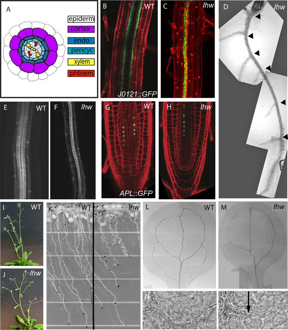
Figure 1. Phenotype of LONESOME HIGHWAY mutants.
(A) Cross section diagram of a mature Arabidopsis root; tissues are arranged radially from outside in: epidermis (white), cortex (light purple), endodermis (dark blue) and stele. The stele consists of a ring of pericycle cells (light blue), surrounding xylem (yellow) and phloem (red) arranged in bilateral symmetry. (B–C) Confocal images of wild type (B) and lhw-1 (C) expressing xylem-associated pericycle marker J0121::GFP in green. Roots are counterstained with propidum iodide (PI, red) to visualize outlines of cells. (D) Brightfield image of lhw-1 root with all lateral roots (black arrowheads) emerging from single side of primary root. (E–F) Confocal image of basic fucshin staining of xylem in wildtype (E) and lhw-1 (F). (G–H) Confocal images of wild type (G) and lhw-1 (H) expressing phloem marker APLpro::APL-GFP in green. (I–J) Whole plant phenotypes of WT Col (I) and lhw SALK_079402 (J). (K) Root growth of wild type (left) and lhw-1 (right) on agar plates at 20-dpg. Note root waving and short root phenotypes in lhw-1. (L–M) vascular pattern in mature (13 dpg) WT (L) and lhw-1 (M) cotyledons (N–O) higher magnification images of xylem from images L and M, respectively. Black arrow points to end of mature xylem elements, to the right, elongated cells typical of procambium are still seen. For each marker, the WT and lhw image pair are at same magnification.

