Figure 4.

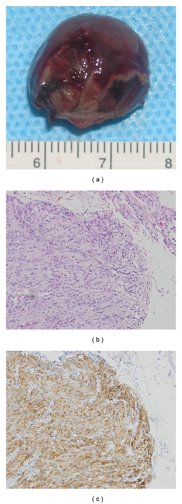
(a) Macroscopic view. Resected tumor was 18∗15∗15 mm in diameter without injury of pseudocapsule. (b) HE staining revealed that spindle cells were proliferating in the submucosal layer. Mitosis was seen less than three cells/50 HPF. (c) The majority of the tumor cell was positive for immunostaining of KIT.
