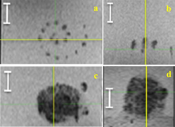
Figure 4.
The top view (a) and the side view (b) of the spiral scan without ADV or acoustic trench. Note the sparse lesioning. In comparison, the top bottom view (c) and the side view (d) of the spiral with ADV and acoustic trench, and with the same individual lesion positioning. Note the complete coverage of the large volume. The white segment is 1 cm. The yellow line shows the plane of intersection with the orthogonal view.

