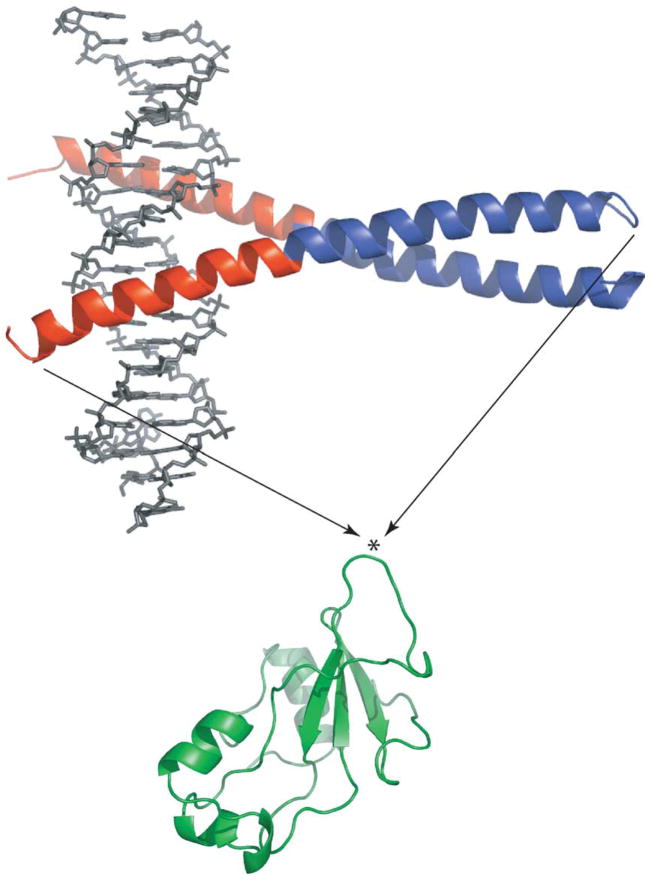
Figure 1.
Creation of the BG fusion protein from GCN4 (top) and Bn (bottom). The DNA binding and coiled-coil regions of GCN4 are colored red and blue, respectively. The bound DNA oligonucleotide is shown in grey. The asterisk indicates the point where GCN4 was inserted (between residues 66 and 67 of Bn). KpnI and NheI restriction sites were created to fuse the Bn and GCN4 genes. The extra nucleotides introduced Gly-Thr and Ala-Ser at the junction points. These dipeptides serve as short linkers. Images were generated by the Pymol program (DeLano Scientific).

