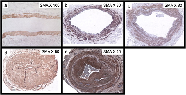
Fig. 2.
Histopathology of vein specimens from normal patients to stenosed AVF. (a) Shows normal human vein in a patient with no CKD. Note the absence of medial thickness and neointimal hyperplasia (b–d) shows SMA sections of patients with advanced CKD at the time of AV access placement. Note that neointimal hyperplasia in patients is variable from minimal neointimal hyperplasia to very severe lesions (e) shows a human vein in a patient with ESRD with a stenotic AV fistula. Note the aggressive thickening of the neointima and media and significant luminal stenosis that is similar to the lesion prior to access placement in some patients (d).

