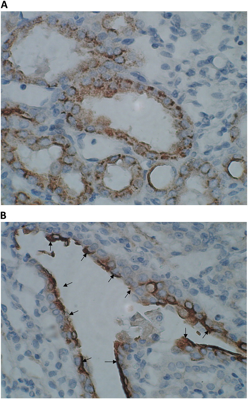
Fig. 8.

Expression of OPN in kidneys of rats that consumed HLP and produced CaOx crystal deposits. Crystal deposition caused tissue disruption. Tubules with as well as without crystals showed heavy expression of OPN. (A) Expression of OPN in cortical tubules. OPN is present both on the luminal surface of epithelium as well as inside the cells. (B) Surface of the urothelium covering papillary surface was heavily stained. The picture shows expression of OPN on urothelial surface in the renal fornix.
