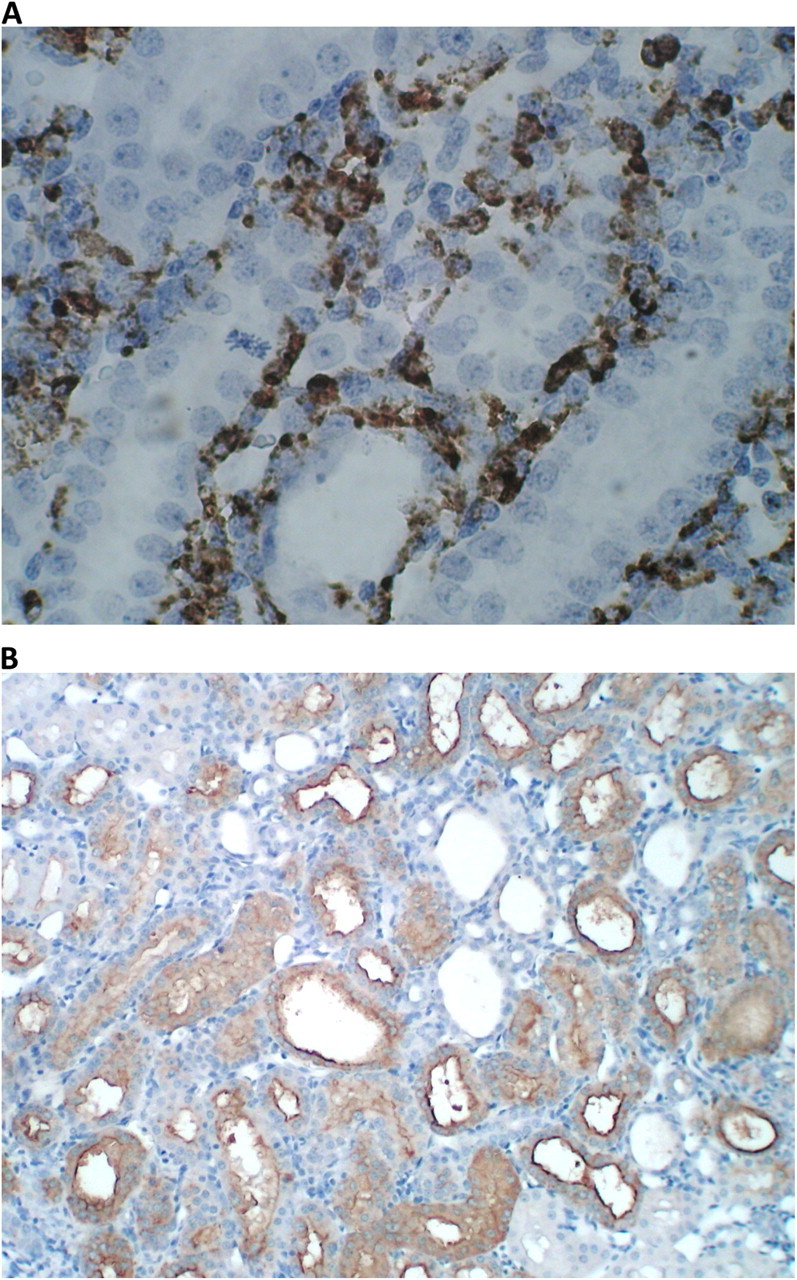
Fig. 9.

Expression of ED-1 and KIM-1 in kidneys of rats that consumed HLP and produced CaOx crystal deposits. (A) Heavy ED-1 staining around renal tubules in the cortex. The picture shows areas with intact tubular epithelium. (B) Heavy expression of KIM-1 in renal cortex. Tubules are dilated and their epithelia are heavily stained. Intertubular area is invaded by inflammatory cells.
