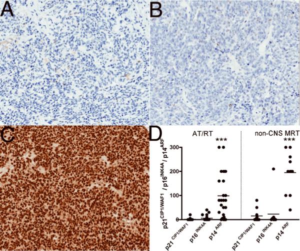
Figure 3.
Immunohistochemical expression profiles of CDK4 and cyclinD1 in atypical teratoid/rhabdoid (AT/RT) and non-CNS malignant rhabdoid tumors (MRT). (A) CDK4 immunostaining showed weak (1+) expression in AT/RT. (B) Semiquantitative estimation of CDK4 shows weak expression in both AT/RT and non-CNS MRT. (C) Focal weak (1+) expression of cyclin D1 staining in AT/RT. (D) Semiquantitative estimation of cyclinD1 showed focal weak expression in both AT/RT and non-CNS MRT. The y-axes in B and D represent a semiquantitative measure of the product of staining intensity and percentage of positive cells in each case.

