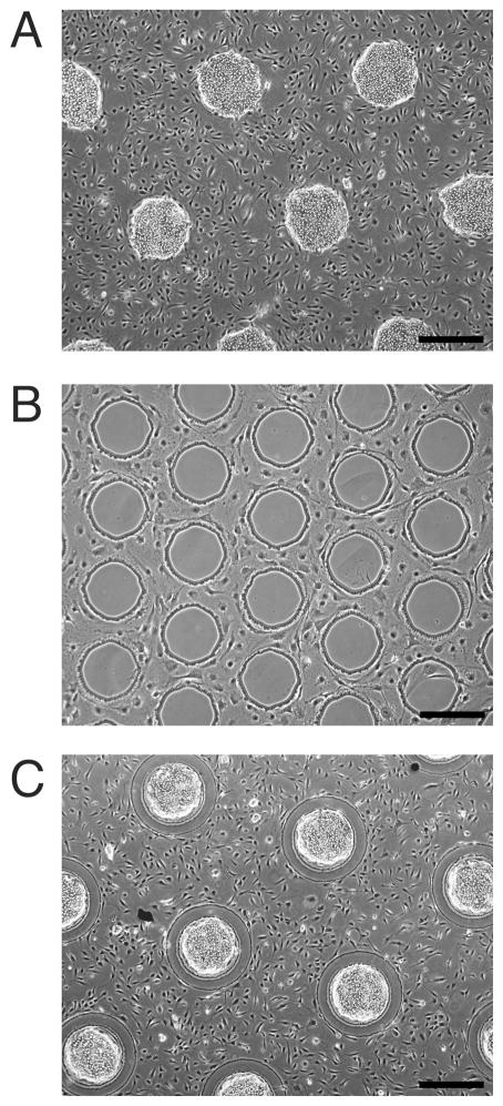
Fig. 2. Control of cell organization via patterned surface chemistry.
(A) Phase contrast image of hepatocytes selectively adhered to islands of collagen, with 3T3-J2 fibroblasts filling the remaining bare glass regions (day 2). (B) 3T3-J2 fibroblasts excluded from islands of PEG-disilane, (day 5). (C) Hepatocytes and 3T3-J2 fibroblasts patterned on a combination of collagen and PEG-disilane, (day 2). Scale bars 500 μm.

