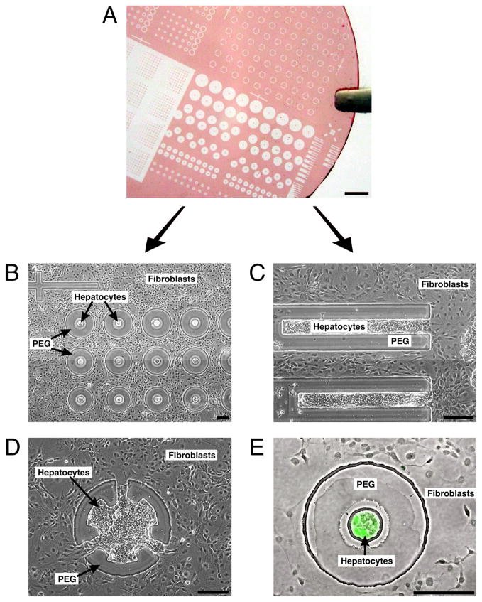
Fig. 3. Parallel screening of multiple cell configurations.
(A) Glass coverslip with patterned photoresist (PEG mask) illustrating the variety of designs possible on a single substrate. Scale bar is 2 mm. (B) Phase contrast image showing variations in hepatocyte-fibroblast separation (day 6). (C and D) Spatially restricted contact between hepatocytes and fibroblasts (day 6). (E) Phase contrast and fluorescence overlay showing expression of intracellular albumin in green after 8 days of culture, indicating retained hepatocyte function. This in situ assay identifies an interesting experimental condition that can be further pursued in larger formats. (B-E) Scale bars are 200 μm.

