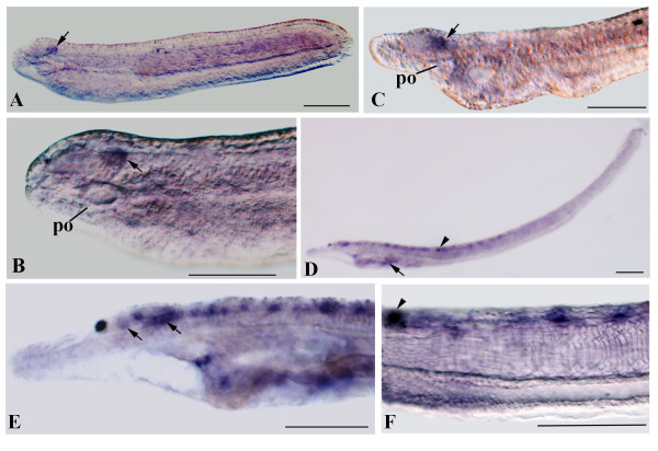
Figure 7.
miR-137 expression in B. floridae. A: Side view of 24-h larva. B: Anterior enlargement of the specimen in A showing expression in a discrete region of the posterior cerebral vesicle just above the preoral pit (po). C: Higher magnification of the anterior end of a 30-h larva. D: Side view of a three gill slit larva with expression in the neural tube and gill slits (arrow). E: Anterior of larva in D showing expression in different cells of cerebral vesicle (arrows) and in segmentally arranged cells of the neural tube just posterior. F: Enlargement of the posterior neural tube just posterior to the pigment of the first dorsal ocellus (arrowhead) with miR-137 transcripts in cells segmentally arranged in the neural tube.

