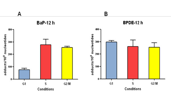
Figure 2.
DNA adduct levels in synchronised MCF-7 cells. Cell were synchronised in G0/G1, S and G2/M phases with different methods, after which they were exposed to 2.5 μM BaP or 0.5 μM BPDE for 12 h. DNA was isolated with a standard phenol/chloroform method and DNA adducts were assessed by the 32P-postlabelling method. Results were expressed as DNA adducts/108 nucleotides.

