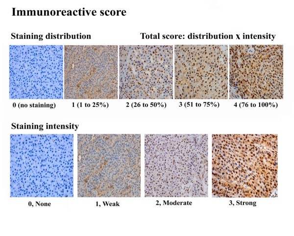
Figure 1.
Typical scored immunohistochemical staining of liver tissue specimens. Formalin-fixed and paraffin-embedded tissues were incubated with primary anti-ZBTB20 polyclonal antibodies (diluted 1:350) at 4°C overnight. The visualization signal was developed with diaminobenzidine (DAB) and the slides were counterstained in hematoxylin. Total score was calculated from sub-scores of staining distribution (0-4) and intensity (0-3).

