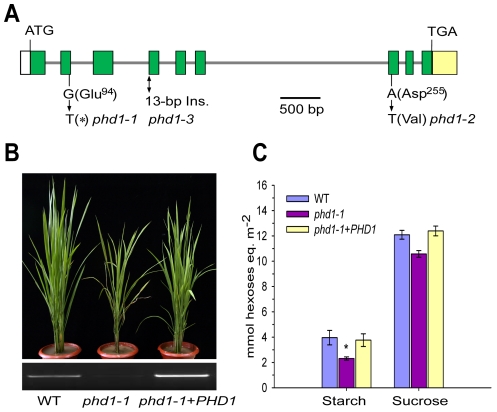
Figure 2. Molecular identification of PHD1.
(A) Structure of the PHD1 gene and its mutation sites in three phd1 alleles. The PHD1 gene consists of nine exons (green boxes) and eight introns (gray lines). Nucleotide insertion and substitutions in the three phd1 alleles are indicated. (B, C) Functional complementation of the phd1 mutant. (B) Upper panel: Phenotypes of wild type, phd1-1, and complemented phd1-1+PHD1 plants at the tillering stage. Lower panel: Expression levels of PHD1 transcripts as detected by semi-quantitative RT-PCR. (C) Sucrose and starch content in flag leaves of wild type, phd1-1, and complemented phd1-1+PHD1 plants at noon of the day at the anthesis stage. Error bars represent SD of eight different individuals. *significant difference between phd1-1 mutant and wild type (P = 0.05).

