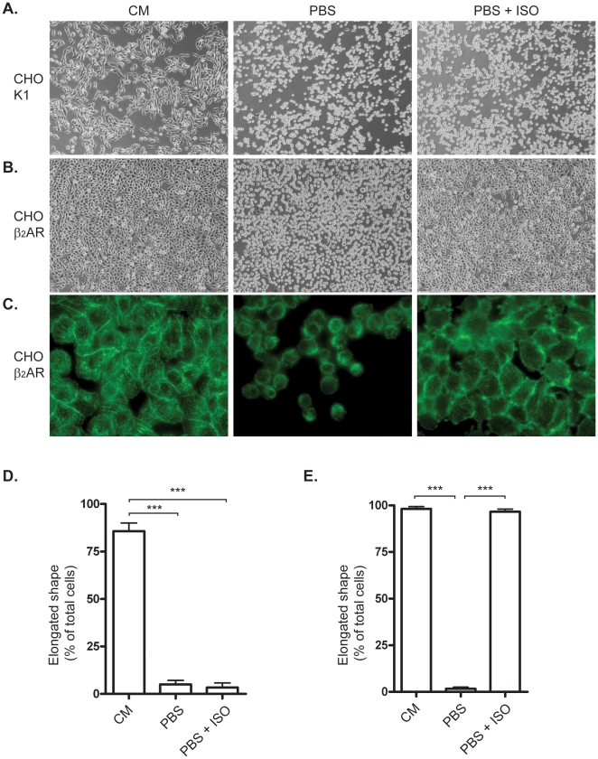
Figure 1. β2-adrenoceptor-mediated inhibition of cell detachment in CHO cells.
A) CHO-K1 cells were kept in culture media (CM) or phosphate buffered saline (PBS) for 20 min with or without 30 min of pre-treatment with 1 µM isoprenaline (ISO). B) CHO cells stably transfected with human β2-adrenoceptor in CM, PBS or PBS+ISO. C) FITC-Phalloidin-stained CHO β2-AR cells in CM, PBS or PBS+ISO. D) Morphology of CHO-K1 cells described by counting cells with elongated shape. The bars represents percentage of total cells. E) Morphology Quantification of CHO-β2 cells. Histograms show percentage of total cells expressed as mean ±SEM of three independent experiments. *** p<0.001, one-way ANOVA.

