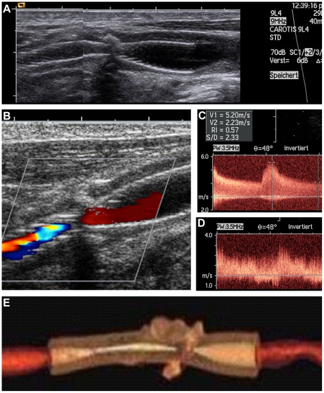
Figure 1. In-stent restenosis after carotid artery stenting as diagnosed during routine duplex sonography follow-up.
Duplex sonography (B-mode) of a carotid artery after stenting showing a narrowing in the middle part of the stent due to a calcified plaque (A). During the routine follow-up investigation after six months there was a typical aliasing phenomenon indicating focal flow acceleration (B). Peak systolic velocity reached up to 520 cm/s (C) with a markedly disturbed poststenotic frequency pattern (D). The reconstructed contrast enhanced computer tomography confirmed the high-grade in-stent restenosis (E).

