Fig. 4.
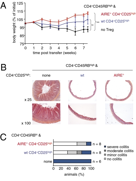
AIRE° CD4+CD25high Treg do not show any defect in prevention of colitis. Colitis was induced as in Fig. 3. (A) Evolution of weight of animals. Shown is the mean weight ± SD as a percentage of weight at the start of the experiment (*P < 0.05, **P < 0.01, Mann–Whitney test; n = 6 without Treg, n = 8 with WT Treg, n = 8 with AIRE° Treg; two independent experiments). (B) Mice were euthanized 7 wk after injection of T cells. Microscopic sections of distal colon were stained with hematoxylin and eosin and examined for histological signs of colitis. Shown results are representative of those obtained in two independent experiments. (C) Colons of mice were examined as in B and clinical scores of colitis were attributed (n values as indicated, from two independent experiments).

