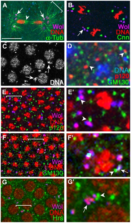
Figure 1. Wolbachia bacteria are present in Golgi-related vesicles.
Wolbachia-infected CS embryos were used to generate all images except (D). (D) was obtained with Wolbachia-free CS embryos. Wolbachia in all images except (A) were visualized with anti-Vang antisera, while those in (A) were visualized with anti-Sdt antisera. Wolbachia, recognized by antisera (blue) and DNA marker (red), appear as pink. In these images, structures appeared as blue do not contain DNA and should be considered to have endogenous Sdt (A) or Vang (B-G except D). DNAs in A, B, C and G were visualized with propidium iodide, and DNAs in D, E, and F were visualized with Toplo-3. (A) In a preblastoderm stage embryo, Wolbachia vesicles (arrows) are attached near the minus ends of the astral microtubules (bracket) but not the polar microtubules (arrowhead). (B) A blastoderm stage embryo shows Wolbachia localization near centrosome (arrow). (C) Wolbachia vesicles are perinulclear during interphase (arrows). (D) GM130 and p120 are present in separate vesicles in Wolbachia-free CS embryos during mid-cellularization. They sometimes are present in the two adjacent vesicles (arrowheads). (E,F) In Wolbachia-infected CS embryos during mid-cellularization, p120-containing vesicles are physically separated from Wolbachia vesicles, but are in proximity (arrowheads) (E). Wolbachia vesicles either are juxtaposed to GM130-containing vesicles (arrowheads) or contain GM130 proteins (arrow) (F). (G) In Wolbachia-infected CS embryos, some Wolbachia vesicles are in proximity with Hrs vesicles (arrow), but did not contain Hrs (arrowhead). Portions marked with brackets in E, F, and G are magnified in E', F' and G'. Scale bar: A,C,E,F,G, 10 µm; B, 4.4 µm; D,E',F',G', 2.5 µm.We then examined the pattern of GM130 and p120 in Wolbachia-infected CS embryos. The Wolbachia vesicles rarely contained p120 protein (<1%; 2/228), but 46% of Wolbachia vesicles located close to the p120 vesicles (106/228) (Figure 1E and 1E'). In contrast, 17% of Wolbachia vesicles contained GM130 protein (40/238), and 76% were juxtaposed to GM130-containing vesicles (180/238) (Figure 1F and 1F'). These data suggest that Wolbachia bacteria reside in a type of Golgi vesicles that are closely related to cis-Golgi. We also found that Wolbachia were not present in endosomes, using an antibody against Hepatocyte growth factor-regulated tyrosine kinase substrate (Hrs) that is present in endosomes [25] (Figure 1G). In conjunction with the previous report that Wolbachia are absent in mitochondria [26], we concluded that Wolbachia are present in a group of cis-Golgi related vesicles.

