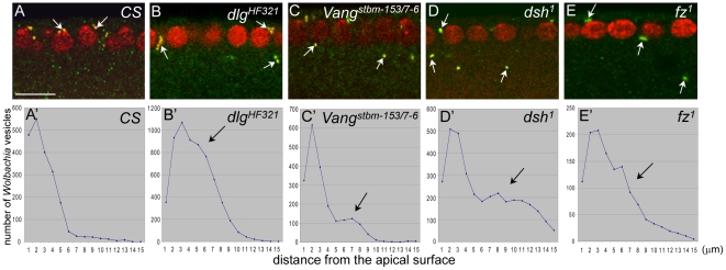
Figure 3. Apical positioning of Wolbachia vesicles is disrupted in dlg, Vang, dsh and fz mutant embryos during early to mid cellularization.
Wolbachia were visualized with anti-Vang antisera and propidium iodide. (A–E) Wolbachia vesicles are marked with arrows. Note that some Wolbachia vesicles in the mutant embryos are present in the basal location (B–E) compared to CS embryo (A). (A'–E') The numbers of Wolbachia vesicles counted with Image J program were plotted. Arrows indicate the region where abnormally high level of Wolbachia along the apical-basal position.

