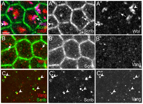
Figure 4. Scrib is present in vesicles that are juxtaposed to Wolbachia vesicles.
(A) In the cortex of cellularizing Wolbachia-infected embryos, Scrib is present in both cell boundary and cytoplasmic vesicles. Scrib vesicles either contain Wolbachia (arrowhead) or are surrounded by Wolbachia vesicles (arrow). Wolbachia were identified with anti-Vang antisera (blue) and propidium iodide (red). Therefore, Wolbachia appear as pink, while endogenous Vang appears as blue in (A). In A”, the strong staining (arrowheads) indicates Wolbachia, but the weak staining indicates endogenous Vang (arrow). (B) In the cortex of cellularizing Wolbachia-free embryos, Vang and Scrib are present in vesicles that are sometimes juxtaposed (arrows). (C) In the embryo interior of cellularing Wolbachia-free embryos, large-sized Vang-containing vesicles all contain Scrib (arrowheads). Scale bar: 3.3 µm.

