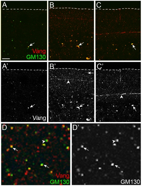
Figure 5. Vang and GM130 are present in same vesicles or juxtaposed vesicles.
(A, B, C) Vesicles containing Vang and GM130 in immediately after egglaying (A), during mid-cellularization (B), and late cellularization (C) are indicated with arrows. Arrowheads in B' and C' indicate the furrow front where Vang is enriched. (D) A tangential section was obtained around 3.5 µm from the apical surface of the embryo in (B) (white line). Arrows indicate the vesicles containing both Vang and GM130, and the arrowhead indicates two juxtaposed vesicles. Scale bar: A–C, 10 µm; D, 2.5 µm.

