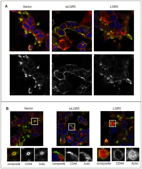
Figure 9. Distribution of CD44 in cells with altered levels of LGR5.
Lim 1899 cells expressing siRNA to LGR5 (siLGR5) or the pTune/LGR5 construct (LGR5) were seeded on chamber slides, fixed and stained with rhodamine-phalloidin (red) or anti-CD44 followed by Alexa 488 anti-rat (green). Nuclei were stained with DAPI (blue). A): typical patterns of CD44 localization in the different cells. B) micrographs selected to show the focal actin rings and their coincidence (vector, LGR5) or lack of coincidence (siLGR5) with CD44. The lower panels show enlarged areas of the micrograph, highlighting the actin structures associated with CD44. In these panels actin staining and CD44 staining are shown separately in greyscale.

