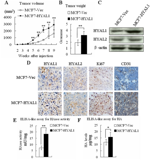
Figure 7. Upregulation of HYAL1 increased tumor growth, angiogenesis, HAase activity and HA expression of MCF7 cells in vivo.
Representative mouse bearing tumors, the average tumor volume (A) from 5th weeks and tumor weight (B) in MCF7-HYAL1 group were increased significantly than in MCF7-Vec group (p<0.01). Western blot (C) and immunohistochemistry (D) analysis showed that the expression of HYAL1 protein was enhanced in the MCF7-HYAL1 group obviously, but HYAL2 levels were not altered. Compared with MCF7-Vec, expression of ki67 and CD31 were increased in MCF7-HYAL1 (D). ELISA-like assay measured HAase activity (E) and HA levels (F) present in the tissue extracts, the HAase activity and HA levels in MCF7-HYAL1 were higher than MCF7-Vec (p<0.01, p<0.05, respectively).

