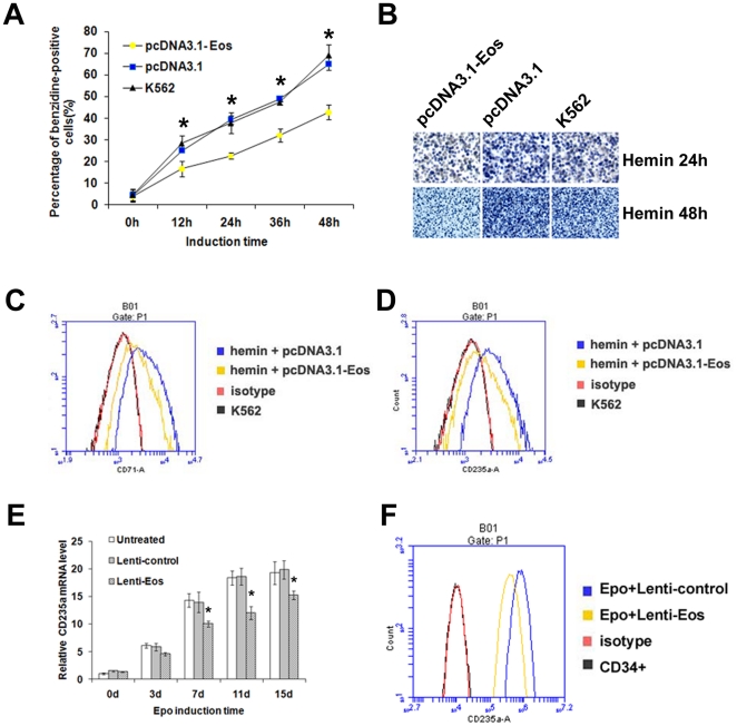
Figure 8. Overexpression of Eos inhibits erythroid differentiation of K562 cells and CD34+ HPCs.
(A) Benzidine staining of K562 cells that were untransfected, transfected with the control vector (pcDNA3.1), or transfected with the Eos overexpression vector (pcDNA3.1-Eos). The percentage of benzidine-positive cells in each group was counted following hemin induction periods of 0, 12, 24, 36, or 48 h. Data were obtained from three independent experiments, and error bars represent standard deviation. *P<0.05. (B) Representative benzidine staining of untransfected and transfected K562 cells that were hemin-induced for 24 or 48 h. (C–D) Flow cytometric analysis of K562 cells transfected with control or pcDNA-Eos and treated with (C) anti-transferrin receptor (CD71) or (D) anti-glycophorin A (CD235a) antibodies after 48 h of hemin induction. (E) Quantitative real-time PCR analysis of CD235a mRNA level during Epo-induced erythroid differentiation of CD34+ HPCs. Error bars represent one standard deviation. *P<0.05. (F) Flow cytometric analysis of CD235a in K562 cells transfected with lenti-Eos or lenti-control after Epo induction for 15 d.

