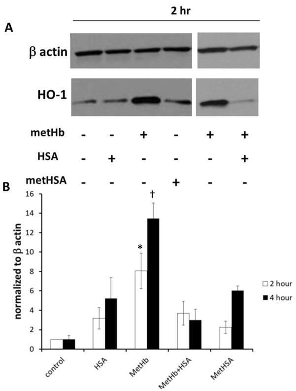
Fig 5.
MetHSA formation inhibits haem oxygenase 1 (HO-1) induction. 100 μM MetHb was incubated in the presence or absence of 600 μM HSA for 4 h at 37 °C. These treatments were diluted 10-fold with serum-free medium and added to BAECs for 2 h or 4 h or cells were treated with synthesized metHSA for 2 h or 4 h. At the end of treatment, complete medium replaced treatment media and cells were collected 24 h later. A) Western blot analysis demonstrating HO-1 expression in BAECs after 2 h treatment. B) HO-1 expression in BAECs after 2h or 4h treatment normalized to β actin (n= 3–11). p<0.05; * metHb+HSA and metHSA at 2 h, †different from all other values at 4 h.

