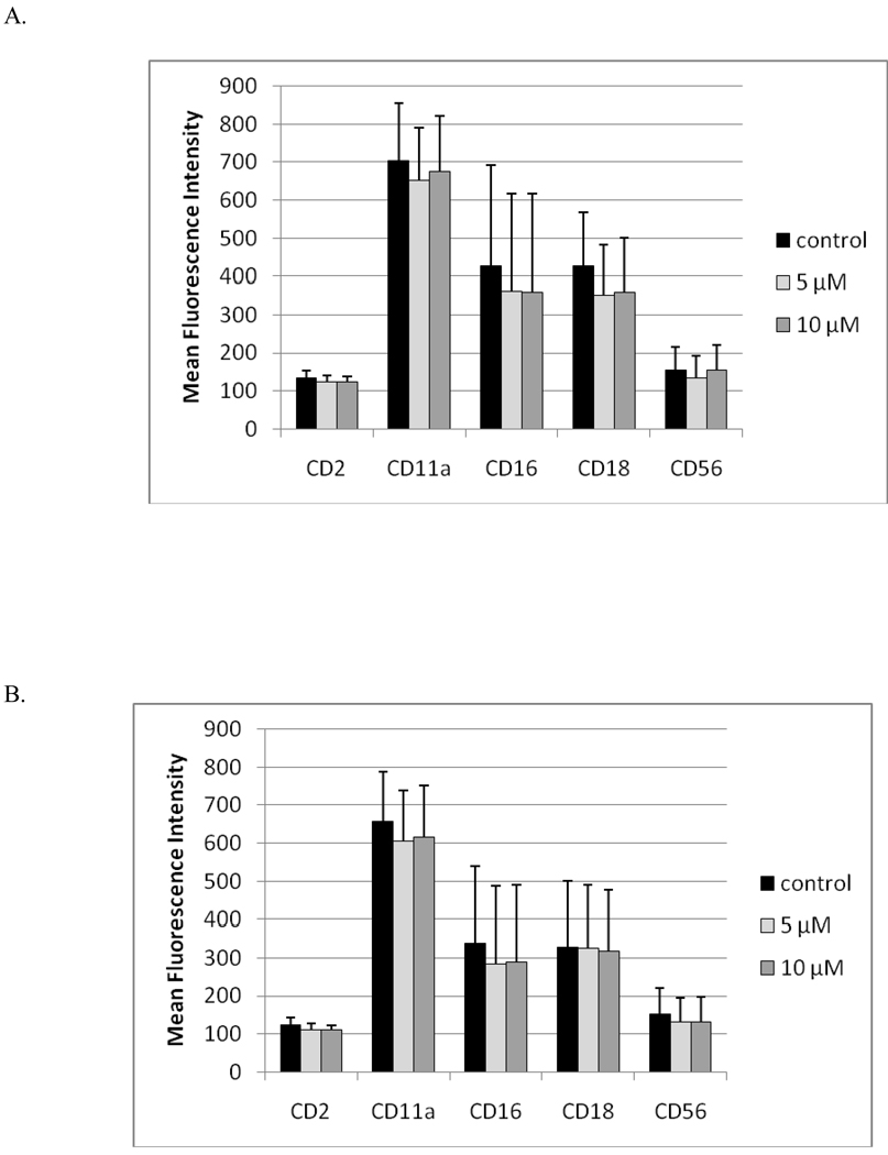
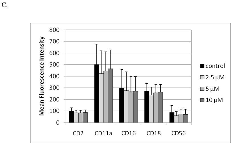
Figure 7. Effects of 1 hr exposures to TBBPA - followed by 24 hr, 48 hr, or 6 d incubations in TBBPA-free media - on NK cell surface protein expression.
(A) 1 hr exposures to 10 µM and 5 µM TBBPA followed by 24 hr incubation in TBBPA-free media. (B) 1 hr exposures to 10 µM and 5 µM TBBPA followed by 48 hr incubation in TBBPA-free media. (C) 1 hr exposures to 10 µM, 5 µM, and 2.5 µM TBBPA followed by 6 d incubation in TBBPA-free media. Values shown are the mean± (± SD) of mean fluorescence intensity (MFI) combined from three different experiments each using a different donor (n = 3).


