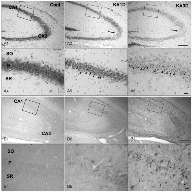
Fig. 1.
Cresyl violet staining (A) and inhibitor of DNA binding 4 (ID4) immunoreactivity (IR) (B) in control (A1, A4, B1, B4) and kainic acid (KA)-treated mice on days 1 (A2, A5, B2, B5) and 3 (A3, A6, B3, B6). (A) In contrast to normal mice tissue, pyramidal degeneration was apparent in the CA3 region on days 1 and 3 post-lesion (arrows in A2, A3). In addition to pyramidal cell loss, small glial cell reactivities were evident (arrowheads in A5, A6). (B) In the control, ID4 IR was not found in the hippocampus (B1). In the KA-injured hippocampus, strong ID4 IR was observed on day 1 post-lesion (B2) and became maximal on day 3 (B3). Higher magnification of rectangular areas (B1-3) in the hippocampus shows sequential changes in ID4 IR cells. Note that ID4 IR cells exhibited the characteristic star-shaped morphology of astrocytes (B6). Cont, control; SO, stratum oriens; P, pyramidal cell; SR, stratum radiatum. Scale bars=200 µm (A1-3, B1-3), 20 µm (A4-6, B4-6).

