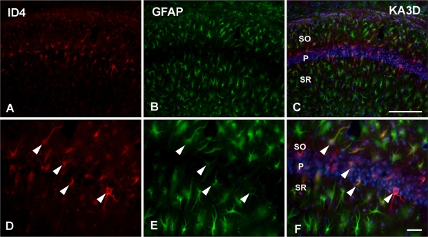
Fig. 2.
Double immunofluorscence staining for identification of inhibitor of DNA binding 4 (ID4)-positive cells in kainic acid (KA)-treated mice. ID4 (A, D) and GFAP (B, E) co-localized well in the CA1 region of the KA-injected hippocampus on day 3 (arrowheads in D, E). GFAP, glial fibrillar acidic protein; SO, stratum oriens; P, pyramidal cell; SR, stratum radiatum. Scale bars=100 µm (A-C), 20 µm (D-F).

