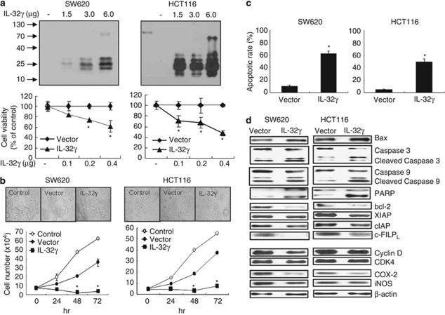
Figure 4.
Expression of IL-32γ, growth rates and apoptotic cell death in IL-32γ-transfected colon cancer cells. (a) Colon cancer cells (1 × 10⁶) were transfected with various amounts of the IL-32γ plasmid (1.5–6.0 μg) for 24 h and harvested. IL-32γ expression was detected by western blotting using monoclonal antibody KU32-52. To determine the effects of different IL-32γ levels on colon cancer cell growth, the cells were inoculated into 24-well plates (5 × 10⁴ cells per well) and transfected with the IL-32γ plasmid (0.1–0.4 μg per well) for 72 h. Cell growth was measured by direct counting after Trypan blue staining. (b) Colon cancer cells were inoculated into 24-well plates (5 × 10⁴ cells per well) and transfected with the vector or IL-32γ plasmid (0.4 μg per well). At 24, 48 and 72 h post-transfection, the cells were harvested by trypsinization and counted after Trypan blue staining. Control: untransfected cells. (c) Colon cancer cells were transfected with the vector or the IL-32γ for 72 h. Apoptotic cells were examined under a fluorescence microscope after TUNEL staining. The total number of cells in a given area was determined by 4′,6-diamidino-2-phenylindole nuclear staining. The results are expressed as mean±s.d. of three experiments with each experiment performed in triplicate. *P<0.05 compared with the vector-transfected colon cancer cells. (d) Cells were transfected with the vector or the IL-32γ for 24 h. Cell extracts were analyzed by western blotting. Each band is representative of three independent experiments.

