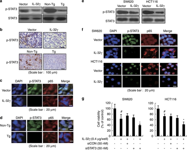
Figure 7.
Effect of IL-32γ on STAT3 activation in tumor tissues and colon cancer cells. (a, b) Phosphorylation of STAT3 in whole extracts of murine tumors, as determined by western blotting (a) and immunohistochemistry (b). (c, d) Cellular localization p-STAT3 (green) and p65 (red) in tumor tissues of xenograft mice (c) and IL-32γ transgenic mice (d). Each image and band is representative of three independent experiments. (e) Colon cancer cells were transfected with the vector or the IL-32γ for 24 h. Whole-cell extracts were prepared and analyzed for phosphorylated STAT3 by western blotting. Each band is representative of three independent experiments. (f) Cellular localization of p-STAT3 (green) and p65 (red) was observed by confocal microscopy after immunofluorescence staining of colon cancer cells transfected with IL-32γ. (g) Colon cancer cells were co-transfected with the IL-32γ and STAT3 siRNA for up to 72 h. Cell growth was measured by direct counting of cells stained with Trypan blue. The results are expressed as mean±s.d. of three experiments with triplicate tests in each experiment. #P<0.05 compared with the colon cancer cells transfected with vector. *P<0.05 compared with the colon cancer cells transfected with IL-32γ alone.

