
Figure 2. Some PF–PC synapses are functional but kinetics and short-term plasticity of PF-evoked EPSC are altered in staggerer mice.
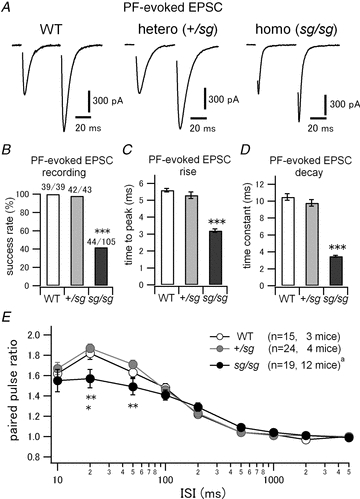
A, EPSCs were evoked by a paired pulse stimulation of PFs at an interval of 50 ms. Stimulus artifacts are blanked. PCs were voltage-clamped at −70 mV. The second EPSCs were facilitated, which is a typical nature of PF–PC synapses. B, rate of successful recordings of PF-evoked EPSCs in WT and staggerer mutants. Each denominator and numerator indicates the total number of tried cells and the number of cells where PF-evoked EPSCs were observed, respectively (WT versus sg/sg and +/sg versus sg/sg, ***P < 0.0001). C, time from stimulus onset to peak of PF-evoked EPSCs (WT versus sg/sg and +/sg versus sg/sg, ***P < 0.0001). D, decay time constants of PF-evoked EPSCs were measured by fitting a single exponential function. (WT versus sg/sg and +/sg versus sg/sg, ***P < 0.0001.) E, mean paired pulse ratios (the second EPSC amplitudes normalized to the first ones) were plotted against varied interstimulus intervals (ISIs) of the two pulses (WT versus sg/sg, *P < 0.05 and +/sg versus+/sg, **P < 0.01). aThe sample size is different at ISI of 2 s (n = 17, 10 mice).