
Figure 1. Synaptically evoked intracellular Ca2+ waves and an associated biphasic membrane potential change in CA1 pyramidal neurons.
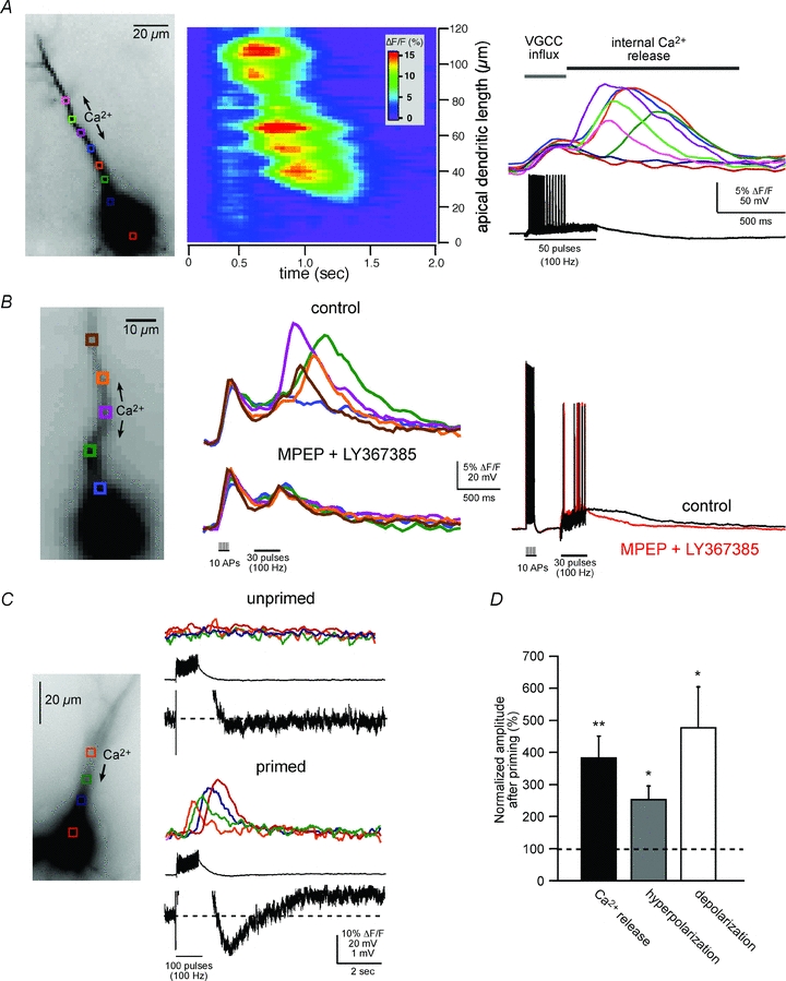
A, left panel, fluorescence image of a fura-2FF-filled neuron. Middle and right panels, two representations of the same imaging data elicited by synaptic stimulation (50 pulses at 100 Hz; see electrical response in right panel). The middle panel is a pseudo-linescan showing that intracellular Ca2+ waves propagate through hot and cold spots of release. The right panel shows coloured waveforms corresponding to Ca2+ rises occurring at the colour-coded boxes (regions of interest, ROIs) over the cell in the left panel. Note the difference in magnitude and timing of the VGCC-mediated Ca2+ rise occurring during the synaptically elicited action potentials and the delayed intracellular Ca2+ wave. B, Group I mGluR blockers selectively blocked intracellular Ca2+ waves, but not VGCC-mediated rises in [Ca2+]i. Rises in [Ca2+]i were first initiated by activation of VGCCs with current injection-evoked spikes (2 ms, 2 nA, 10 spikes at 100 Hz) followed by synaptic stimulation (30 pulses at 100 Hz). Middle panel, mGluR antagonists, MPEP (10 μm) and LY367385 (100 μm), blocked synaptically elicited internal Ca2+ release. Right panel, the evoked electrical waveforms recorded during the Ca2+ responses shown in the middle panel. Note the suppression of the membrane depolarization following bath application of the mGluR antagonists. C, synaptically elicited intracellular Ca2+ waves correlate with a biphasic membrane potential change. Synaptic stimulation, subthreshold for eliciting a rise in [Ca2+]i (100 pulses at 100 Hz), failed to elicit a biphasic membrane potential change. 30 s after ‘priming’ the cell with a train of current injection-evoked spikes (2 ms, 2 nA current injection; 100 spikes at 100 Hz) and consequent VGCC-mediated Ca2+ influx (not shown), the previously subthreshold synaptic stimulation elicited internal Ca2+ release and a hyperpolarization and depolarization. Note that the fast EPSPs evoked by electrical stimulation were not affected by priming. In this example, mAChRs and GABABRs were blocked (1 μm atropine and 1 μm CGP55845, respectively). D, summary data showing priming-induced facilitation of synaptically elicited internal Ca2+ release and associated membrane potential changes (normalized to pre-priming response averages; n = 7; *P < 0.01, **P < 0.001; ANOVA).