
Figure 2. mGluR agonists elicit a Ca2+ wave-dependent hyperpolarization and depolarization.
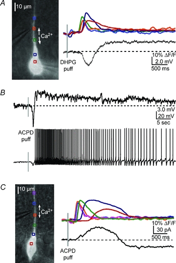
A, left, overlay of a DIC image and fluorescence neuron image showing the position of the pressure application pipette near the primary apical dendrite of the recorded cell. Right, a DHPG puff (400 μm, 50 ms) onto the primary apical dendrite triggered a bidirectionally propagating Ca2+ wave and associated hyperpolarization and depolarization of a CA1 pyramidal neuron. B, upper panel, in a different neuron, an ACPD puff (400 μm, 50 ms) elicited a Ca2+ wave (not shown) and a transient hyperpolarization and a sustained depolarization. Lower panel, when the neuron was held at a membrane potential slightly subthreshold for spiking (∼−53 mV), ACPD elicited a transient hyperpolarization and a sustained train of action potentials. C, in voltage clamp, an ACPD puff elicited an outward current and inward current (see Results for summary data).