
Figure 3. The hyperpolarization and depolarization are due to SK channels and CAN channels, respectively.
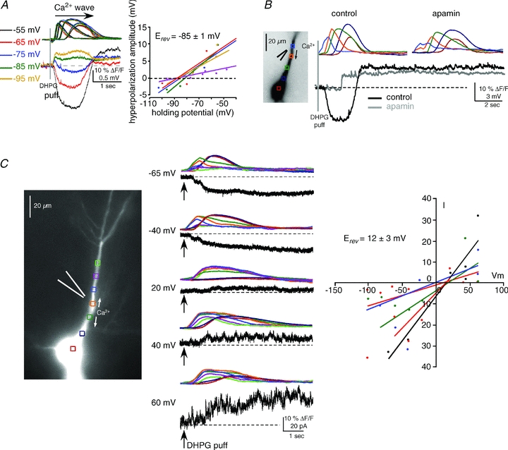
A, left, the reversal potential (Erev) of the hyperpolarizing potential was determined to be ∼−85 mV by applying DHPG puffs (400 μm, 50 ms) at different holding potentials in current clamp and measuring membrane potential changes. Intracellular Ca2+ waves were similar at all holding potentials (waves are colour-coded by holding potential). Right panel, summary graph showing the reversal potential for all cells tested (n = 5; each cell is represented by a different colour). B, consistent with a mechanism involving SK channels, apamin (100 nm) blocked the hyperpolarization. This treatment unveiled the isolated depolarizing potential and revealed its delayed onset. C, left and middle panels, the mGluR-mediated, Ca2+-dependent depolarizing current was isolated in voltage clamp and its Erev was determined to be ∼12 mV. DHPG puffs (400 μm, 50 ms) were delivered to the primary apical dendrite in the presence of voltage-gated K+ channel and Na+ channel blockers, and GABABR blockers (see Results). Right, summary I–V graph for all cells tested (n = 5; each cell is represented by a different colour) shows data consistent with activation of CAN channels.