
Figure 6. TRPC1, TRPC4 and TRPC5 antibodies block the mGluR-mediated and intracellular Ca2+ wave-dependent depolarization.
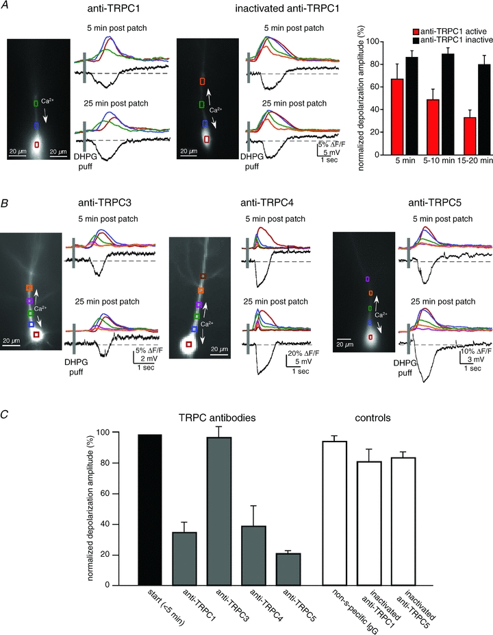
Antibodies to TRPC were loaded into patch recording pipettes (1:100 dilution). In some cases antibodies were heat inactivated. Responses recorded ∼5 min after breaking into the cell were compared to responses recorded ∼20 min after breaking in. A, an example of data collected from a CA1 pyramidal neuron loaded with anti-TRPC1 and an example of a neuron loaded with heat-inactivated anti-TRPC1. Anti-TRPC1 selectively blocked the sustained depolarization. B, examples of neurons loaded with anti-TRPC3, anti-TRPC4 or anti-TRPC5. TRPC3 did not affect the depolarization (n = 3, P > 0.1, t test); TRPC4 (n = 5), like TRPC1 (n = 5) and TRPC5 (n = 5; data not shown), suppressed the mGluR/IP3R evoked-depolarization (P < 0.01 for each antibody, t test). C, summary data for anti-TRPCs, and the controls, IgG (n = 7) or inactivated anti-TRPC1 (n = 5) or TRPC5 (n = 5).