
Figure 8. TRPC1, 4 and 5 antibodies suppress mGluR-mediated increases in spike frequency.
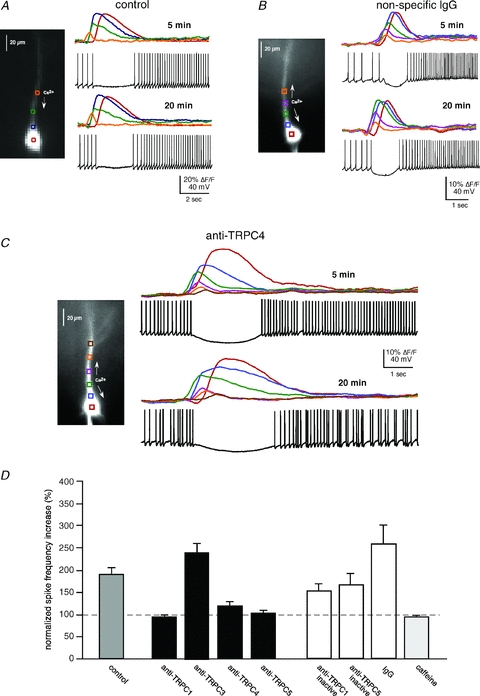
A, mGluR activation of intracellular Ca2+ waves modulates the firing pattern of CA1 pyramidal neurons. Puffing DHPG (50 ms) onto the apical dendrite of a spiking pyramidal neuron held at ∼−45 mV in current clamp suppressed and then increased the firing frequency of this representative pyramidal neuron. B, an example of a neuron loaded with IgG. mGluR regulation of firing frequency was not affected by IgG (or inactivated anti-TRPCs; not shown). C, an example of a neuron loaded with anti-TRPC4. Addition of anti-TRPC1, 4 or 5 (1:100 dilution) to the patch pipette suppressed the increase in firing frequency 20 min after breaking into the cell. D, summary data showing that anti-TRPC1, 4 and 5 suppress mGluR-mediated increases in firing frequency (cumulatively P < 0.01, t test). TRPC3 (n = 3) did not alter mGluR-mediated regulation of firing (P > 0.1, t test). Inactivated anti-TRPCs (n = 4) and IgG (n = 7) did not have a significant effect on mGluR-mediated increases in firing rate (P > 0.1, t test). Caffeine puffs (50 mm; n = 5), which activate an RyR-mediated ISK but not an ITRPC, did not elicit an increase in firing frequency (P > 0.1, t test).