Figure 1.

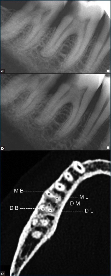
(a) Preoperative diagnostic radiograph; (b) diagnostic radiograph taken from a mesial angulation; and (c) spiral CT scan of the involved tooth showing the presence of 3 distal and 2 mesial canals

(a) Preoperative diagnostic radiograph; (b) diagnostic radiograph taken from a mesial angulation; and (c) spiral CT scan of the involved tooth showing the presence of 3 distal and 2 mesial canals