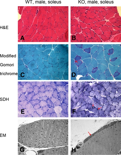
Fig. 2.
Abnormal mitochondrial accumulation in TR4−/− mice muscle. Histological examination of frozen soleus muscle tissues from 1-yr-old TR4−/− and TR4+/+ mice (A–F). The cryostat sections were subjected to several staining procedures including H&E staining (A and B), Trichrome staining (C and D), and SDH staining (E and F). The typical ragged red fibers, which represent higher staining activities of mitochondria, are indicated by red arrows. The photos were taken under ×400 magnification. G and H, Electron microscopy (EM) examination of soleus muscle from 1-yr-old TR4−/− and TR4+/+ male mice. The abnormal subsarcolemmal aggregates of mitochondria in TR4−/− samples are indicated by red arrows. The soleus muscle tissues from the TR4−/− and TR4+/+ mice were fixed, embedded, and examined by electron microscopy. Examinations were carried out in three pairs of wild-type (WT) and TR4 knockout (KO) mice, and representative pictures are shown.

