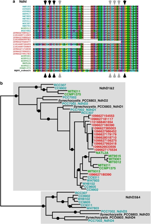
Figure 4.
Viral NAD(P)H dehydrogenase subunits. (a) NdhI protein alignment. Synechococcus sequences are colored in cyan, Prochlorococcus in green and viral proteins in red. The Nqo9 sequence from T. thermophilus is shown for reference. The conserved cysteine residues coordinating Fe–S clusters in Nqo9 are marked with arrows (black for N6a and gray for N6b). For clarity, only part of the protein length is shown. (b) NdhD FastTree approximated maximum-likelihood phylogenetic tree. Synechococcus NdhD sequences are colored in cyan, Prochlorococcus in green and viral proteins in red. Synechocystis PCC6803 and Synechococcus PCC7002 NdhD1, 2, 3 and 4 sequences were used as references. Only bootstrap values above 80% are shown as black circles on the branches.

