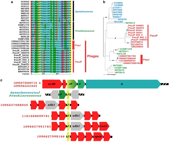
Figure 5.
Viral PSI PsaJ protein. (a) PSI PsaJ protein and peptide alignment. Viral PsaJ proteins identified in this study and viral PsaJF (only the N-terminus, which contains the PsaJ portion, is shown) are labeled in red to the right of the alignment. (b) PsaJ FastTree approximated maximum-likelihood phylogenetic tree. Synechococcus PsaJ sequences are colored in cyan, Prochlorococcus in green, viral PsaJF and the newly identified viral PsaJ in red. Only bootstrap values above 80% are shown as black circles on the branches. (c) Schematic physical maps of selected viral clones or Prochlorococcus and Synechococcus genome fragments containing the PSI psaJ gene and a viral GOS clone containing the psaJF fusion gene. ndhI denotes NAD(P)H dehydrogenase I gene, speD a polyamine biosynthesis gene, talC a transaldolase gene and nrdB the ribonucleoside-diphoshate reductase-β 2 gene. Red-arrowed boxes mark viral genes and gray mark bacterial genes. PSI genes are colored in yellow, green and blue.

