
Fig. 1.
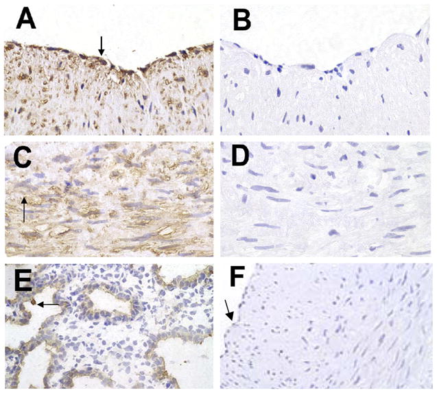
Surfactant protein D (SP-D) in human coronary arteries (HCAs). Paraffin sections of HCAs were immunostained for SP-D and then counterstained with hematoxylin. Positive staining was detected in endothelial cells (ECs; A, arrow) and medial smooth muscle cells (SMCs; C, arrow). B and D: staining controls in which PBS was used in place of the primary antibody. E: positive control of human fetal lung explant tissue immunostained for SP-D protein; the arrow points to type II ECs. F: control stained with nonimmune IgG; the arrow points to the EC layer. Images are representative of sections from 4 different specimens (all images were photographed at ×40 and enlarged similarly).