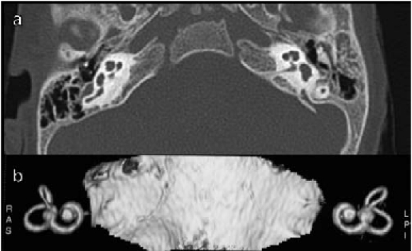
Fig. 3.

a) CT shows the anomalous anatomy of the patient. There is bilateral agenesis of lateral semicircular canal, no identifiable bone layer between basal and second turn of the cochlea and a narrowed internal auditory canal; b) same anomalies at MRI 3D reconstruction.
