
Fig. 1.
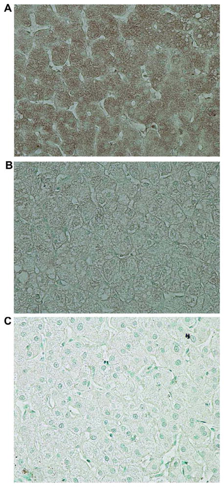
Immunohistochemistry of SOCS3 in patients with chronic hepatitis C. (A) Typical staining in a non-responder. Strong SOCS3 staining is seen in hepatocyte cytoplasm. (B) Typical staining in a responder. SOCS3 is faintly stained in hepatocyte cytoplasm. (C) Negative control using normal rabbit serum in a non-responder. Original magnification 400×. [This figure appears in colour on the web.]