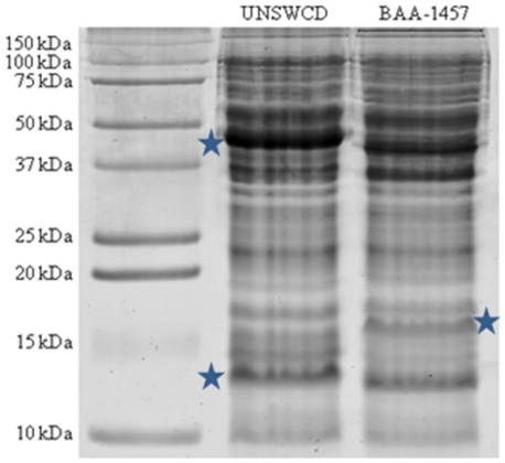
Figure 5. One-dimensional polyacrylamide gel electrophoresis of whole cell lysates of C. concisus UNSWCD and BAA-1457.
Each gel lane for each strain was sectioned into 25 gel slices and processed for mass spectrometry analysis. Regions within the gel labeled with stars correspond to areas reflecting high diversity between the protein profiles of the two strains.

